
Dhealthwellness.com -The index of suspicion for gout diagnosis is low, both clinically and radiologically. This was highlighted by McCracken et al. in 1986, and this remains true today. This is due to the nonspecific nature of the disease, which is often undiagnosed on imaging. According to Golden (1956), only about one-third of advanced gout cases show sufficient radiological evidence to be diagnosed. The radiologist can use advanced imaging techniques to detect early gout and determine the severity of the disease.
A Good Choice for Diagnosis and Treatment of Gout
During the first 6 to 8 years after the onset of gout, radiographic changes are not present. The acute stage of gout may only present with asymmetric soft tissue swelling, which resolves in a few months. However, new evidence supports the use of advanced ultrasound techniques to detect occult destructive arthropathy and early bone erosions in patients with gout. Further, if an individual has a history of gout, a specialized MRI is an excellent option for the diagnosis and treatment of this disease.
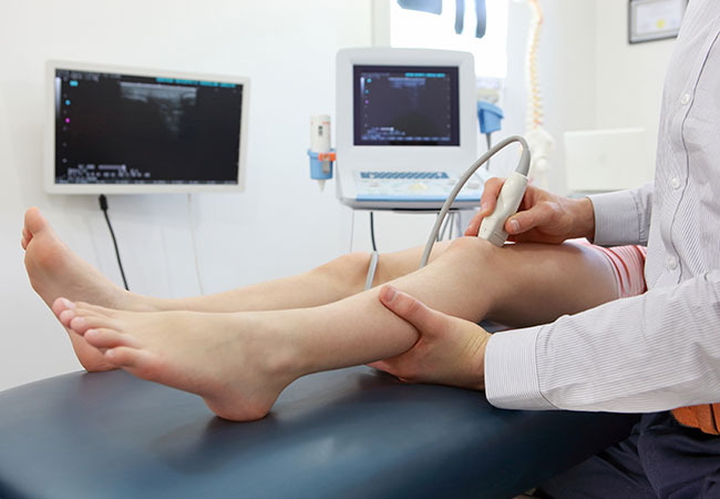
An ultrasonographic study is also beneficial in determining if the disease is responding to treatment. This type of imaging is particularly useful in cases of atypical presentations, including young adults, older patients, or subcutaneous tophi. For instance, an ultrasound study performed in conjunction with polarizing microscopy can determine whether a patient has an underlying inflammatory disease such as gout. In addition, a recent review article in the journal Radiology described the utility of computerized tomography in gout.
The gold standard in the diagnosis of gout is the analysis of synovial fluid, but this is an invasive procedure. With the help of ultrasound, the doctor can diagnose gout more accurately than with conventional CT. In the US, the physician can evaluate tophi by hand without exposing patients to ionizing radiation. The US can also guide a doctor to aspirate tophus and joints. The results of the US can aid in determining the type of treatment required.
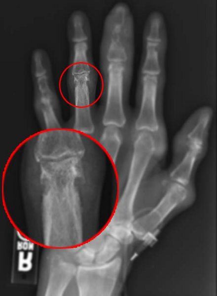
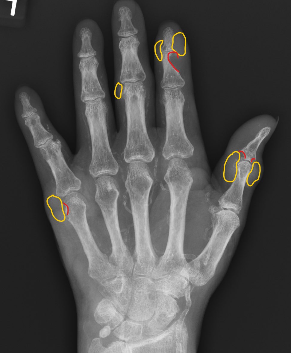
Radiological Findings in Gout
The most common radiological finding in gout is a bone erosion, accompanied by calcification of the joints. The patient may also exhibit soft-tissue masses. The most common types of ultrasound examination are MRI, CT scans of the knee and ankle, and plain radiography of the elbow. These images can help the physician determine the type of treatment for gout and its risk factors. Despite the various imaging techniques available, they are not fool-proof.
Although conventional radiography is the preferred method for diagnosis of gout, CT is a specialized diagnostic tool. It can give detailed images and reveal bone erosions. The CT can also reveal a number of other conditions, including calcification and inflammation. Unlike conventional radiography, CT can be very helpful in the early stages of gout. But it can also be risky. Some people are not candidates for CT.
The diagnostic tools used in gout include ultrasonography and CT. This type of imaging allows the doctor to evaluate gout with ease. In contrast, the MRI is a diagnostic tool that can reveal the presence of gout. It is also useful in diagnosing the disease with ultrasound. If you think you may have gout, you should visit your doctor immediately. It is the only way to be certain that you are free of the condition.
Essential Diagnostic Tool for Gout
Ultrasound is an important diagnostic tool for gout. Its sensitivity and accuracy are greater than those of conventional X-ray. High-resolution ultrasound can be a more efficient alternative than a conventional X-ray for diagnosing gout. For example, a high-resolution CT can show a tophi with a higher level of resolution than a traditional X-ray. Its sensitivity also allows the physician to view the crystalline structure of the affected joint.
Although X-rays are the most common diagnostic tool for gout, they have low sensitivity and specificity. Compared to the high-resolution US, X-rays are not the only diagnostic tools for gout. Moreover, they take six to twelve years to find. In other words, MRI is a non-invasive method that can be used for evaluating the tophi of the patient. It is also a very important tool in the evaluation of gout.
The most common gout MRI is an invasive test that uses a needle. This method is best for evaluating patients with tophaceous tophi. It is also very useful for assessing the extent of tophaceous deposits in the affected joint. In addition, it is helpful in monitoring treatment for gout. There are many new drugs for gout that have improved its treatment options. This allows doctors to monitor the effectiveness of the drug.
Reference: