Dhealthwellness.com – Symptoms of Ulnar Styloid Process Pain can include pain in the hand and wrist, as well as the thumb. It is often related to a tear in the Triangular Fibrocartilage Complex, which is a part of the ligaments that hold your fingers together. It is also associated with Distal Radioulnar Joint Instability, a condition that involves instability of the joint. It is possible to treat this condition through surgical intervention.
Symptomatic Ulnar Styloid Fracture Treatment is Surgery
Traditionally, treatment of symptomatic ulnar styloid fractures has been a surgical excision. However, two studies have suggested that transosseous tissue repair may be a suitable alternative in cases where the ulnar styloid is nonunion or non-union with an adjacent distal radial fracture. For symptomatic ulnar styloid base fractures with a small avulsion bone fragment, suture anchors or tension band wires are a possible option. In the present study, surgical repair of 31 patients of unilateral ulnar styloid fractures with distal radioulnar instability was performed between 2004 and 2017. These patients had a mean follow-up of 2.2 years.
Patients were divided into two groups. The first group consisted of nine patients who underwent open reduction and plate fixation. The second group consisted of 21 patients who underwent tension band wire fixation. Both groups showed similar rates of surgical complications. Implant-related complications were significantly more frequent in the tension band wire group. The incidence of nonunion was also higher in the tension band wire group.

Symptoms of Ulnar Styloid Process are pain, swelling and bruising along the ulnar side of the wrist. This is caused by impaction between the ulnar styloid process and the triquetrum. During the impaction, the triangular fibrocartilage complex (TFCC) becomes irritated and wears down. Eventually, it tears and the bones are pressed against each other. This friction can cause painful synovitis.
Ulnar Styloid Fracture Causes Wrist Pain
Ulnar styloid fractures are relatively common. They often occur as a result of a fall onto an outstretched hand. In addition to causing wrist pain, they may also cause additional injury to the wrist ligaments. These injuries usually require surgery. A splint is placed over the wrist to prevent movement. It is usually made of plaster or fiberglass. In severe cases, metal pins may be placed to secure the bone. The wrist is immobilized for two weeks. Then, the bones are aligned by a surgeon.
The surgeon may need to remove part of the styloid process or resew it. He may also insert pins, an external fixator, or a plate and screws to secure the bones. This surgical procedure is called a styloidectomy. TFCC tears are very common injuries that occur in the wrist. They are often the most neglected high-energy injury of the wrist joint. They can occur with minor trauma or as a result of degenerative changes in the wrist.

In many cases, TFCC tears are caused by degenerative conditions that lead to the degeneration of the fibrocartilage in the wrist. In some cases, a degenerative tear is caused by repetitive gripping. It is also possible for a TFCC tear to occur in a younger patient. Surgical treatment for a TFCC tear may be necessary. Surgery may improve the patient’s pain and ability to perform certain activities. However, surgery should only be considered after other methods have been tried. Surgical treatment may include casts or splints.
Non Surgical Treatment of a TFCC Tear
Nonsurgical treatment of a TFCC tear may include physical therapy, rest, and casts. Surgery may be considered after the patient has been treated with nonsurgical treatment for several months. The type of treatment is dependent on the patient’s age, the extent of the damage, and the cause of the injury.
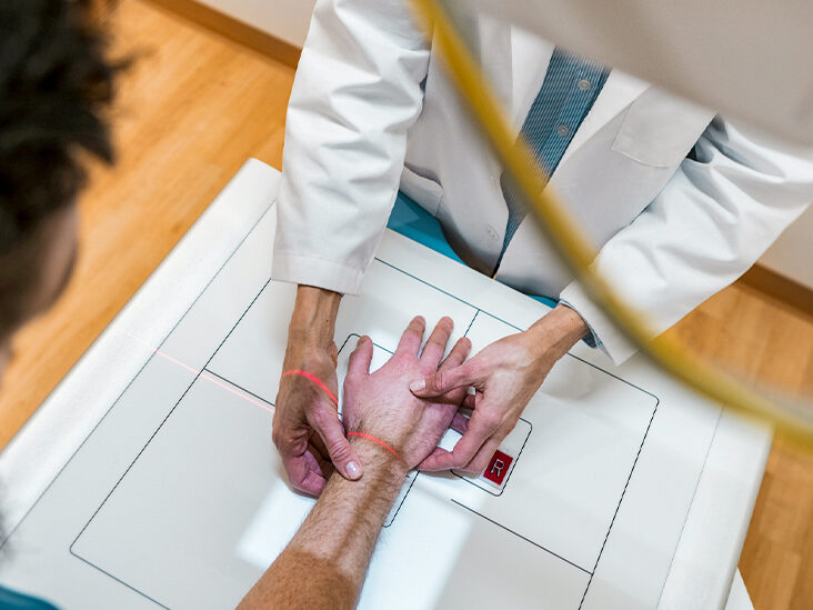
Several imaging modalities are available to determine the extent of distal radioulnar joint instability. Computed tomography (CT) and magnetic resonance imaging (MRI) are valuable tools. A CT diagnostic criteria has been proposed for DRUJ instability. The Mino method involves a line drawn through the ulnar border and the chord of the sigmoid notch. This method is also known as the congruency method. Computed tomography (CT) scans obtained in extreme pronation or supination will provide additional information regarding DRUJ instability. A CT scan obtained in extreme pronation will show the ulnar styloid process and ulnar head as well as the triangular fibrocartilage complex (TFCC).

Computed tomography (CT) scanners are valuable tools for evaluating DRUJ integrity. This is especially true in cases of acute distal radioulnar joint instability. Surgical repair may be performed under general anesthesia. This may include open reduction, TFCC examination, and possible repair. The ulnar styloid fracture is commonly associated with distal radius fractures. This type of fracture disrupts the attachment of the TFC and can result in progressive damage to the TFCC. The fracture may also impinge on the extensor carpi ulnaris tendon sheath. In addition to this, traumatic dislocations can occur.
Reference:
