
Dhealthwellness.com – A 24-year-old woman is diagnosed with osteonecrosis of the sesamoid bone. She experiences increasing pain since last August. Her pain is described as a small cramp when walking, but she cannot recall any fall or bad jump. She does not drink alcohol or run much, so she cannot attribute the pain to another factor. She is also unable to drive, despite being diagnosed with osteonecrosis of the sesamoid.
Causes of Sesamoid Bone Osteonecrosis
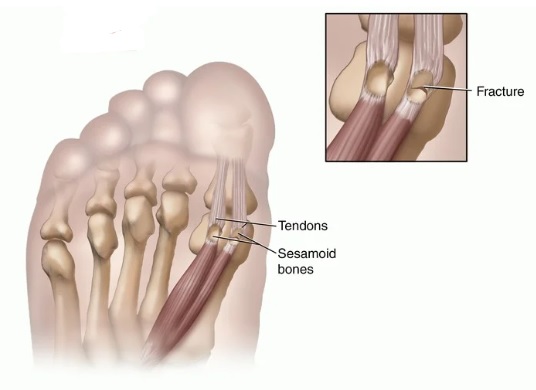
Sesamoid bones are small, nodular foci made of bone and cartilage. There are about 42 sesamoid bones in a person. They are located in the big toe joint and are linked to tendons and muscles. Sesamoid bones allow the big toe to move normally and absorb pressure from the ball of the foot. However, overuse of the sesamoid bones can lead to osteonecrosis of the sesamoid bone. For this reason, exercise routines should be moderately intense at first and increase in intensity over time. Alternating between different types of exercises is also recommended to protect the sesamoid bones.
Osteonecrosis of the sesamoid bone is rare and can cause significant limitations. Resection of the sesamoid is often considered the treatment for the condition, but resection of the affected sesamoid has its own set of limitations. A patient with this condition was treated conservatively and returned to sports activities without any surgery. The treatment for sesamoid osteonecrosis is determined by the severity of the symptoms and the severity of the pain.
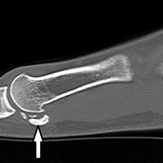
Avascular osteonecrosis of the sesamoid is a rare but dangerous condition that occurs as a result of repetitive trauma. While the etiology is unclear, some authors postulate that the condition may be related to repeated trauma, disrupting blood supply. However, there are some cases in which avascular osteonecrosis of the sesamoid does not develop in these circumstances.
Sesamoid Osteonecrosis Treatment
The treatment of sesamoid osteonecrosis has limited success and often requires surgery. Fortunately, radial extracorporeal shock wave therapy is a novel conservative treatment for osteonecrosis of the sesamoid bone. In this case, a 52-year-old club tennis player presented with a 1-year history of pain in the left forefoot, aggravated with walking and recreational tennis. Further, she had no prior history of any injury that had caused the condition.
Surgical treatment of osteonecrosis of the sesamoid bone involves an incision in the bottom of the foot. The doctor then separates the soft tissue surrounding the sesamoid bone. The incision is then closed. Recovery of the affected foot may take anywhere from three to six months. If treatment does not work, a bone graft may be necessary. A limb-saving surgery may also be necessary.
While osteonecrosis of sesamoid bone is not a common condition, the disease can be debilitating and severely limit a person’s activity. Osteonecrosis of sesamoid is a condition that can be fatal if left untreated. The symptoms of osteonecrose of sesamoid bone are not easy to identify, but proper treatment can alleviate pain and minimize the risk of bone fracture.
Diagnosis when Experiencing Signs of Foot Pain
A 24-year-old female patient is a goalkeeper on a college futsal team. She experienced right foot pain for two months after futsal training sessions. The pain gradually grew worse, until she discontinued futsal. Direct palpation of both sesamoid bones showed pain that was reproduced by direct pressure over the fabella. The pain was more severe on extension of the knee and was reproducible when a person applies direct pressure to the sesamoid bone.
 Because avascular necrosis causes bone death, there is no blood supply left. The symptoms include a deep ache in the foot, a painful joint, and the surface of the bone collapses. The condition can be accompanied by severe arthritis of the foot. To assess the extent of osteonecrosis, doctors may suggest physical therapy or surgical treatment. Sometimes, joint replacements are required.
Reference: