
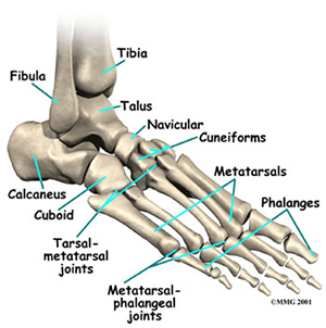
Dhealthwellness.com – The first and fifth metatarsals form the proximal metatarsal joint. These condyloid joints have shallow cavities and elliptical surfaces. They are supported by two collateral ligaments and allow for flexion, abduction, and circumduction. The third and fourth metatarsals are connected at the base of the toes. The proximal metatarsal joint is also known as the second toe.
Types of Injury to the Proximal Metatarsal Joint
The proximal metatarsal joint is located in the proximal foot. The first metatarsal joint rotates about its helical axis. This is the only fracture in which the axes are oriented. It is important to note that the axis of the joint is influenced by the age of the patient. The proximal metatarsal is the most common type of injury, but fractures of any part of the joint can be problematic.
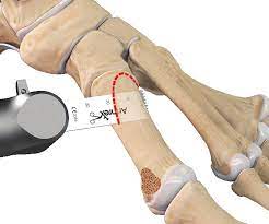
MRI studies of proximal fifth metatarsal fractures present important diagnostic challenges. A millimeter difference can lead to a vastly different treatment regimen and prognosis. Patients with suboptimal treatment can suffer delayed union, reinjury, and chronic disability. The difficulty is compounded by the confusion surrounding the term “facial” and “metatarsal.”
There are many types of fractures of the proximal metatarsal joint, but one is described as a type of osteochondral fracture. The asymmetric angulation of this joint is a key factor in metatarsophalangeal joints. Because the anatomy of the proximal metatarsals is so variable, this asymmetry can lead to a wide range of symptoms and diagnoses.
Common Proximal Fifth Metatarsal Joint
The proximal fifth metatarsal joint is the most common of the metatarsal bones in the foot. Its location makes it important to determine the type of fracture. A lateral longitudinal arch includes a tuberosity, while the medial longitudinal arch has a cuboid and a metatarsal 4 and 5 fracture. Chi Nok Cheung was the first person to describe a lateral long-distance avulsion and a proximal distal articulation.
The proximal fifth metatarsal joint is a complex articulation between the rounded metatarsal heads. The articular surfaces of the first metatarsal head are transversely convex. The proximal fifth metatarsals are mainly vertical. The first apex of the fifth metatarsal is triangular and asymmetric, allowing for a greater degree of plantar flexion and abduction.The proximal metatarsal joint is poorly described in the literature. The joint has variable blood supply between the different parts of the bone. The proximal diaphysis receives blood from multiple metaphyseal vessels while the tuberosity receives blood from a nutrient artery. In fractures of the proximal metatarsal joint, a disproportionate amount of bone is exposed, leading to higher failure rates.
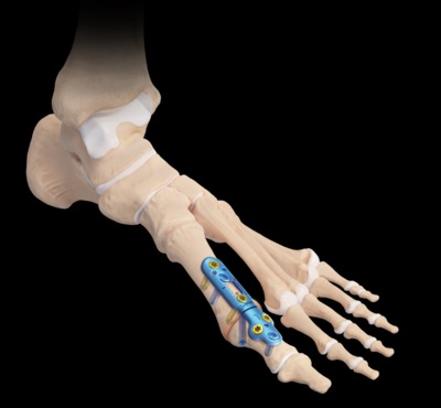
The proximal metatarsal joint is divided into several parts. The first metatarsal is the most common. Its size ranges from 187 to 185 mm2 and is related to arch height and the foot’s structure and function. During exercise, the foot is frequently inverted, causing the metatarsal to rotate and flex. The proximal metatarsal may become dislocated and inflamed.
Affects Foot Position and Arch
The first metatarsal is rotated around the helical axis. For N=9 feet, this rotation occurs at the junction of the metaphysis and the diaphysis. Its rotation is related to the arch height. The orientation of the first metatarsal joint is a vital part of the foot’s structure and function. This rotation affects the position of the foot and the arch.

The fifth metatarsal is the most commonly affected. Its proximal intermetatarsal surface is triangular, and the lateral fourth proximal intermetatarsal are triangular. Approximately 80% of specimens have a triangular articular surface, and the lateral proximal fourth is a monomorphic. The lateral third metatarsal has a triangular articular surface.
Proximal Metatarsal Joint Surface
The proximal metatarsal joint surface typically reflects the distal joint surface of the medial cuneiform. The proximal metatarsal is flat, with two facets separated by a transverse ridge. The fifth metatarsal is a bipartite medial cuneiform. Its shape is 8-shaped. However, it does not display a tripartite medial cuneiface.
The third metatarsal is asymmetric. Its asymmetrical position is called hallux valgus. Its asymmetrical alignment of the metatarsal joint can result in instability of the third metatarsal. If the joint is unstable, it can cause pain and other complications. There are three types of proximal metatarsal joints. These are the first, proximal, and fifth.
Reference: