Dhealthwellness.com – Having Dysplastic Atypical Syndrome can be very frightening for you and your loved ones. It is a rare disorder that causes abnormal growths in the brain, and you will want to be aware of all of the symptoms so you can get the diagnosis and treatment you need. You will also want to know how to prevent the symptoms from occurring.
Comprehensive Screening for Atypical Dysplastic Syndrome
Getting a thorough checkup to find out if you have Dysplastic Atypical Syndrome can be very frightening is not a bad idea. Having the correct information is the best way to avoid having them removed for cosmetic purposes. Depending on your skin type, your doctor may use a local anesthetic to perform the excision. The procedure is usually minor and scarring is limited. Your doctor may also close the wound with sutures. Typically, the sutures are removed soon after the procedure.
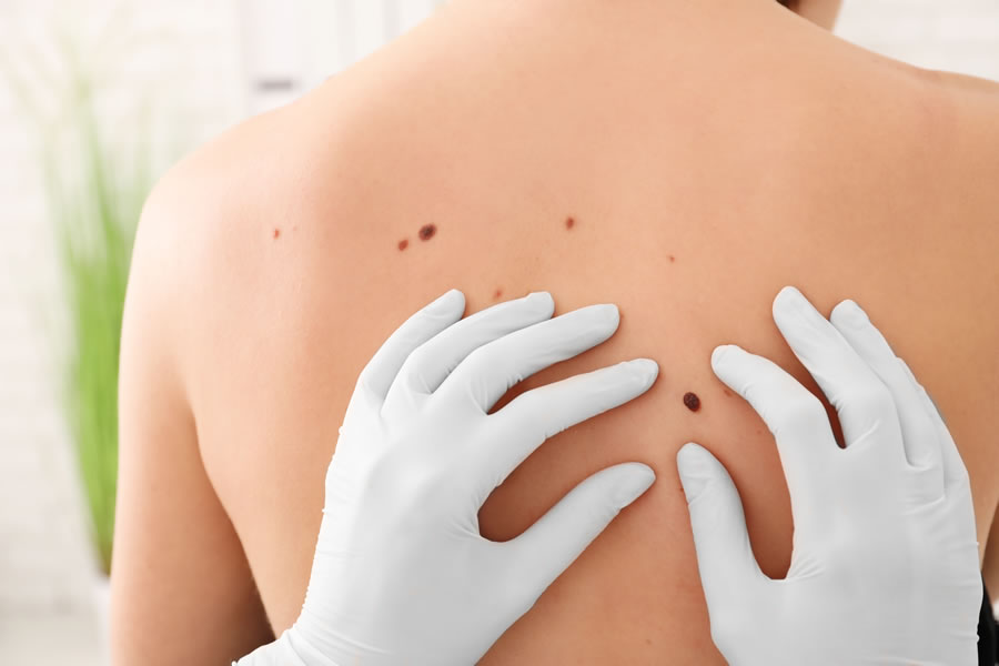
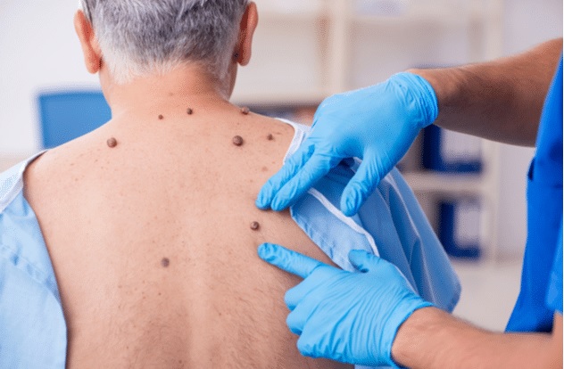
Atypical Moles are benign moles that are found on the chest, back, or any other part of your body that you might have spent a lot of time in the sun. They are not cancerous, but they do increase your risk of developing melanoma. If left untreated, melanoma can spread throughout your body and is extremely dangerous. Fortunately, it is usually curable when detected early. The best way to detect Atypical Moles is to have regular skin checks performed by a licensed medical professional. This is especially important for fair-skinned people.

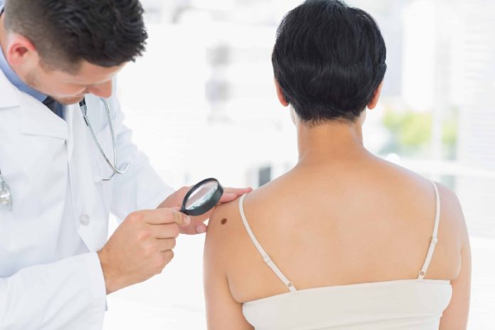
The detection of dysplastic atypical moles is an important step in the diagnosis of melanoma. These lesions are associated with an increased risk of developing melanoma. The best way to detect changes in your skin is to have a regular skin exam with a dermatologist. Atypical moles are often larger than common moles. They can have irregular borders, apebly surfaces, or a mixture of colors. They are usually found on the back, chest, and legs, but can appear on other parts of the body. They can also appear on the scalp.
Dermatologist Performs a Mole Biopsy to Check for Melanoma
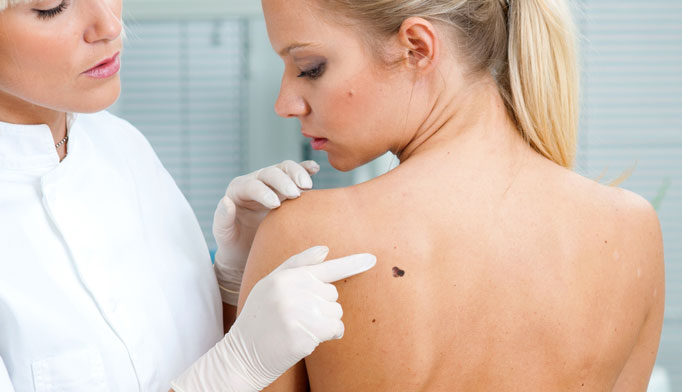
Atypical moles can be removed for cosmetic reasons or if a dermatologist suspects that the mole may be a sign of melanoma. If you have a personal history of skin cancer or if you have a family history of it, see a dermatologist at least once a year. Atypical moles are generally diagnosed on the basis of clinical features. In some cases, a dermatologist may also biopsy the mole to check for melanoma.
Unlike common moles, dysplastic nevi are benign, but they may develop into malignant melanomas. For this reason, regular skin checks are a must. Using a reputable dermatologist to perform a thorough examination is the best way to ensure a healthy future. A typical mole is larger than a quarter, has a smooth or wavy surface, and may change color over time. A typical atypical nevi consists of a tan or brown scaly lesion with an irregular border and a diameter of at least 5 millimeters. There are various shades of coloration and may be on the chest, back, or extremities. In some cases, atypical nevi may occur on the forehead or in the hairline. They may also be accompanied by a cluster of scabs.

Atypical moles can be cured with surgical or non-surgical means and should be monitored closely. As with any skin condition, patients should make sure to wear sunscreen and reduce their exposure to the sun in general. Surgical removal of dysplastic atypical moles may be necessary to prevent melanoma from developing. However, appropriate surgical management of dysplastic atypical lesions is not well defined. In addition to surgical removal, patients with dysplastic atypical lesions should receive regular skin examinations, patient education on melanoma prevention, and skin care.
Atypical Dysplastic Lesions are Characterized by Irregular Borders
Dysplastic atypical lesions are characterized by irregular borders, irregular color, and atypical coloration. They are typically larger than 5 mm in diameter and have a characteristic appearance. They are usually light in color, with variable coloration, and they may have flat or raised areas. They may also appear on the skin of the back, chest, buttocks, and extremities.

Some of these lesions may appear early in life, while others may appear at any age. Atypical nevi have an increased risk of developing cutaneous melanoma. However, melanoma is often curable when it is detected early. The risk of developing melanoma increases with the severity of abnormality.
Reference:
